To contact us Click HERE
Motivation: During patient presentations, saying that a patient is "dizzy" without further qualifiers is sure to trigger further questions. Did dizziness actually mean vertigo or fainting ("presyncope") or unsteadiness? This approach to dissecting dizziness into further subdivisions stems from a 1972 paper which divided dizziness into vertigo, presyncope, disequilibrium, and "vague lightheadedness," with the implication that vertigo stems from vestibular causes, presyncope from cardiovascular causes, disequilibrium from neurological causes, and other dizziness from other causes. This approach, while never rigorously validated, has permeated medicine and has almost become the standard framework for evaluating dizziness. Recently, this approach has been questioned with growing evidence that the various subtypes of dizziness have many overlapping etiologies.
Results: Unlike other posts, I will summarize three articles here that show that the approach of subdividing dizziness may not be valid because many dangerous causes of dizziness have variable presentations.
1. Culic, V., Miric, D. and Eterovic, D. "Correlation between symptomatology and site of acute myocardial infarction." Int. J. Cardiol. (2001) 77: 163-8:
In this paper, the authors attempted to correlate sites of myocardial infarction and symptoms of presentation among 1546 patients. Among the many symptoms, the authors separated feeling of "vertigo" from "faintness." Here are the findings:
SITE OF INFARCTION % WITH VERTIGO % WITH FAINTING
Anterior 11.1 6.2
Inferior 4.7 4.9
Lateral 8.3 4.2
2. Newman-Toker, D.E. and Camargo, C.A. "Cardiogenic Vertigo - true vertigo as the presenting manifestation of primary cardiac disease." Nature Clin. Prac. Neurol. (2006) 2: 167-172.
This is a case report of a 90 year old woman who presented to the ED after saying that "Everything's going around in a circle" followed by a brief period of decreased consciousness without prodromal feelings of palpitations or presyncope. In the ED, presence of vertigo was used to rule out cardiac causes, and patient was admitted to Neurology. Monitoring there revealed periods of transient asystole (14 second pauses) during which the woman had similar feelings of vertigo. Placement of a pacemaker resolved these episodes. This case-report also referred to another study summarized next.
3. Low, PA, Opfer-Gehrking, TL, McPhee, BR, et. al. "Prospective evaluation of clinical characteristics of orthostatic hypotension." Mayo Clin. Proc. (1995) 70: 617-22.
This article examined 90 patients with documented orthostatic-hypotension undergoing tilt-table testing. When patients were put in an upright position, 88% complained of lightheadedness while 37% complained of vertigo with some patients complaining of both symptoms.
Discussion: I thnk that these studies (along with many others) make the point that using the type of dizziness to exclude causes of dizziness is dangerous and can lead to mistakes. Other more validated algorithms have been created (such as using "timing and trigger" by Dr. Newman-Toker), which use the onset of symptoms and factors provoking the symptoms to generate a differential. Also, people experience dizziness in many ways, and the same cause could trigger many symptoms both in the same person and in different people. As far I could tell, there have been no large scale studies which have particularly looked at errors generated by relying on dizziness symptoms. At some level, dizzy is just, well, dizzy.
30 Mayıs 2012 Çarşamba
Improving the CHADS2 Score
To contact us Click HERE
Motivation: Recently, I met a man with CHADS2 score of one. We shook hands, and he had atrial fibrillation. Slowing his heart down was relatively easy, but what about anti-coagulation, aspirin or warfarin? A CHADS2 score of one is a grey "intermediate risk" zone without clear indications for using warfarin or aspirin. About two years ago, a group from England and Netherland proposed a further refinement to the CHADS2 score (called CHA2DS2-VASc) to help decide on anti-coagulation in these intermediate risk cases.
Paper: Lip, G.Y.H., Nieuwlaat, R., Pisters, R., et. al. "Refining Clinical Risk Stratification for Predicting Stroke and Thromboembolism in Atrial Fibrillation Using a Novel Risk Factor-Based Approach: The Euro Heart Survey on Atrial Fibrillation." Chest (2010); 137: 263-272.
Methods: The authors proposed a revised point scoring system with the following components:
Congestive Heart Failure or LV dysfunction: 1
Hypertension: 1
Age 75 or over: 2
Age 65 or over: 1
Diabetes: 1
Stroke, TIA, or other embolic disease: 2
Vascular disease (MI, PAD, or known aortic plaque): 1
Female gender: 1
To validate the predictive value of this scoring system, the authors analyzed data from 1,084 patients without mitral stenosis or heart valve surgery who did not use warfarin. The authors tracked survival status and risk of thromboembolic disease. The patients were derived from Euro Heart Survey cohort, which tracked patients among 182 hospitals in 35 countries.
Results:
Cohort Characteristics: The patients in the tracked cohort were on average 66 years of age and 40.8% were women. The most common risk factor was hypertension (67.3%) followed by CAD (38.4%). Overall, 34.9% of the cohort had CHADS2 score of one, 20.4% with score of zero, and rest with higher scores. 74% took anti-platelet agents.
Thromboembolic Risk: The annual risk of thromboembolic events by risk factor is shown below (only the low risk rates are shown). Adjusted rate for aspirin use assumes a 22% risk reduction by aspirin use.
Score: 0 (103 patients, no risk factors) - Event rate: 0, annual rate adjusted for ASA use: 0
Score: 1 (162 patients) - Event rate: 0.6%, annual rate adjusted for ASA use: 0.7%
Score: 2 (184 patients) - Event rate: 1.6%, annual rate adjusted for ASA use: 1.9%
Score: 3 (203 patients) - Event rate: 3.9%, annual rate adjusted for ASA use: 4.7%
One of the novel risk factors that this scoring system adds is female gender. In univariate analysis, female gender is associated with event rate odds ratio of 2.53 (1.08-5.92).
Discussion: This paper provides help in further distinguishing patients really at low risk not requiring further treatment. I was pretty impressed with the zero annual event rate in patients with no risk factors. On the other hand, for patients with score of 2, treatment with warfarin is likely indicated since the risk of symptomatic embolic complication (1.9%) balances the risk of major bleeding (about the same risk). Especially in an older population, aspirin carries a major bleeding risk (estimated to be about the same in elderly population) but provides substantially decreased protection against embolic disease.
The study, while helpful, has some limitations. First, the analysis was done retrospectively in a cohort. It is unclear why these patients were not anti-coagulated. It is possible that factors which led to decision not to anti-coagulate also substantially modified the subsequent risk of embolic events (high risk behaviors such as IV drug use). Nonetheless, I think that this study adds to the identification of the truly low risk group.
Does venipuncture cause lymphedema for woman after breast cancer surgery?
To contact us Click HERE
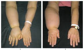
Does venipuncture cause lymphedema for woman after breast cancer surgery?
Motivation: An 85 year old woman is admitted from the ED for IV
antibiotic treatment of community acquired pneumonia. During the
night, the RN pages me because the peripheral IV in the patient’s
right hand infiltrated and attempts to place another IV in that arm
were unsuccessful. A simple solution would be to try on the left hand
but both the RN and the patient are adamantly against this idea
because the patient had “breast cancer surgery” on the left side 20
years ago. They state that inserting a peripheral IV in the left hand
would cause lymphedema which would become debilitating. However, it
seems peculiar to me that having a distant surgery would preclude the
use of an entire extremity therefore I set out to find the evidence.
Studies:
1. Clark B, Sitzia J, Harlow W. Incidence and risk of arm oedema
following treatment for breast cancer: a three-year follow-up study.
QJM. 2005
2. Winge C, Mattiasson AC, Schultz I. After axillary surgery for
breast cancer–is it safe to take blood samples or give intravenous
infusions? J Clin Nurs 2010;19:1270–1274.
3. May;98(5):343-8.Hayes SC, Janda M, Cornish B, Battistutta D, Newman
B. Lymphedema after breast cancer: incidence, risk factors, and effect
on upper body function. J Clin Oncol. 2008 Jul 20;26(21):3536-42.
Methods:
1. Prospective 3 year observational study of 188 woman who had
undergone surgery for breast cancer that involved sanpling, excision
or biopsy of their axillary nodes.
2. Questionnaires were mailed to all 311 patients who had undergone
removal of axilliary nodes in a major Swedish hospitals in 2000-2001.
Study published in 2010 so I assumed 7-8 years of follow up.
3. Observational study of 287 Australian women with invasive breast
cancer were evaluated 6-18 month after treatment.
Results:
1. At follow-up, 39 (20.7%) had developed lymphoedema. Hospital skin
puncture (vs. none) (RR 2.44, 95%CI 1.33-4.47), mastectomy (vs. wide
local excision or lumpectomy) (RR 2.04, 95%CI 1.18-3.54), and BMI > or
= 26 (vs. BMI 19-26) (RR 2.02, 95%CI 1.11-3.68) were the only
significant risk factors.
2. 88 of 311 women reported venipuncture on the affected arm. Only 4
patients developed lymphedema.
3. 33% (n = 62) of the sample were classified as having lymphedema; of
these, 40% had long-term lymphedema. Older age >50, more extensive
surgery (ie, mastectomy), and having a sedentary lifestyle
significantly increased odds (three- to six-fold) of lymphedema.
Removal of 20 or more lymph nodes, experiencing one or more
treatment-related complication or symptom, and being single each
increased odds 2.6- to 5.0-fold, but CIs were wide and included 1.0.
Conversely, having a lower yearly income significantly decreased odds
of lymphedema over the following 12 months five-fold.
Discussion: Lymphedema results when lymphatic drainage systems are
damaged leading to accumulation of interstitial fluid. More than 2
million American women are diagnosed with breast cancer each year with
more than 200,000 developing lymphedema secondary to cancer or its
treatment. Patients with lymphedema suffer chronic pain and swelling
that causes significant physical and mental anguish and leads to
decreased productivity. Treatment for this condition remains
suboptimal therefore attention has focused on prevention.
The above referenced studies all seem to agree that there are certain
risk factors for developing lymphedema including more extensive
surgery and obesity/sedentary life style. Two studies which
specifically looked at venipuncture in the affected arm had
conflicting results. Clark et al. 2005 was a prospective
observational study which provides level 2 evidence linking
venipuncture with increased risk of lymphedema while Winge 2010 was a
retrospective questionnaire study providing only level 3 evidence
against such a linkage. Clearly, the evidence is far from perfect.
Given the importance of the problem, a randomized clinical trial to
study preventive measures for lymphedema should be undertaken.
In the mean time, the results of these studies should motivate us to
attempt to clarify the nature of the patient’s cancer and surgery
before declaring an extremity off limits. Common sense seems to
suggest that we should avoid the affected arm in patients who are
older, obese and have had aggressive disease requiring more extensive
surgery and lymph node dissection. A patient who had DCIS and was
treated with breast conserving therapy without axillary dissection is
less likely to develop lymphedema. In addition, we need to take into
account the clinical context in which this dilemma takes place. For my
patient, a simple external jugular would have probably solved the
issue. On the other hand, in a patient who is having a massive upper
GI bleed and needs every single access that we can provide, the
uncertain risk of future lymphedema is clearly secondary to the
immediate risk of death from hypovolemia.
Lastly, a quick search of the internet will reveal that breast cancer
survivor’s networks and patient advocacy websites regarding lymphedema
are extremely prevalent and vocal about the need to avoid the affect
extremity. Therefore, communication with our patients is essential.
As with any medical procedures, the risks and benefits should be
clearly delineated and the patient included in the decision making
process. In this way, we avoid the anger and frustration that develops
when patients feel their concerns are not addressed, even in the
absence of lymphedema itself.
Pulmonary Embolism and Atrial Fibrillation
To contact us Click HERE
Motivation: Last November, I admitted a patient with dyspnea. He had new onset atrial fibrillation, and discussion in the morning predictably revolved around causes of atrial fibrillation. One of the causes mentioned was pulmonary embolism (the "green book" lists it under pulmonary triggers). I find PE to be a strange trigger though. I associate atrial fibrillation with processes that chronically stretch the atria resulting in a dilated atrium, but PE is an acute process. Searching the literature at that time did not reveal any evidence for or against assoication of atrial fibrillation with PE. Recently, when revisiting the question, I found an article by a European group published in December of 2011 addressing this issue.
Paper: Gex, G., Righini, M., Gal, G.L., et. al. "Is atrial fibrillation associated with pulmonary embolism?" J. of Thrombosis and Haemostasis (2011) [e-pub ahead of print]
Methods: To analyze association, the authors pooled together data from two large trials (CT-EP3 and CT-EP4) investigating PE diagnostic strategies. In both studies, patients were included if they presented with clinical suspicion of PE defined largely as acute dyspnea or chest pain without obvious cause. All patients had EKG at baseline with confirmation of PE diagnosis by CT.
Results:
Cohort: Total of 2,449 patients were analyzed. The mean age was 59.9 years and 43.7% were males. Of this group with clinical suspicion of PE, 22.6% had PE confirmed by CT.
Association with AF: Atrial fibrillation was detected at baseline in 133 patients. Prevalence of PE was 22.8% (n=519) in patients without atrial fibrillation but 18.8% (n=25) in patients with atrial fibrillation (p = 0.28). To adjust for differences in comorbidities between patients with AF and those without AF, the authors next created an adjusted model accounting for age, sex, CHF, COPD, stroke, renal clearance, and neoplasm. After adjustment, there was no increased association with AF and PE (OR of 0.68, 0.42-1.11, p = 0.122). AF failed to be associated with PE even in patient less than age 65 (OR 0.86, 0.35-2.12) or with no heart failure (OR 0.63, 0.37-1.06). Authors attempted to separate association of AF with presenting symptoms (dyspnea or chest pain), but the numbers were too small to make meaningful conclusions.
Discussion: This paper establishes that in patients presenting with acute dyspnea or chest pain, finding atrial fibrillation on EKG does not meaningfully change the likelihood of PE. If anything, finding atrial fibrillation had a trend towards reducing the likelihood of PE (adjusted OR of 0.68, 0.42-1.11). This effect is likely from similar presenting symptoms of atrial fibrillations and PE. While this paper in part answers my initial question of the utility of using atrial fibrillation to suspect PE, there are some important limitations. First, the paper only examined patients presenting with acute dyspnea or chest pain. The paper did not examine patients with new onset AF and ask how many had PE. Therefore, the results cannot be extended to patients with atrial fibrillation without obvious symptoms though presumably the incidence of PE would be even lower. Also, given the structure of the paper in which PE had to be a likely diagnosis, patients with AF with RVR and dyspnea were likely excluded. Incidence of PE in this population is also unclear. Depsite these limitations, I think that this paper provides important insights into the limited utility of considering AF in the diagnosis of PE.
CSF Cortisol in Meningitis
To contact us Click HERE
Motivation: This year, I have seen two patients with community acquired meningitis - both had viral meningitis. When we stopped antibiotics on both, I had slight trepidation about what if we were wrong. After all, microbiologic data on CSF can be misleading. For example, in about 10% of patients, bacterial meningitis can present with lymphocytic predominance. Low plasma glucose is present in only 50-60% of patients with bacterial meningitis. Recently, I came across this article evaluating CSF cortisol as a specific marker of acute bacterial meningitis.
Paper: Holub, M., Beran, O., Dzupova, O., et. al. "Cortisol levels in cerebrospinal fluid correlate with severity and bacterial origin of meningitis." Critical Care (2007) 11:R41
Methods: Study conducted in an academic hospital in Prague. Inclusion criteria were symptoms of menigitis (fever, headache, meningismus) for less than 72 hours and lumbar puncture performed on admission prior to administration of steroids as part of meningitis treatment. Bacterial meningitis was diagnosed by positive bacterial CSF culture or detection of bacterial DNA in CSF using PCR. These patients were compared retrospectively to data from 37 patients with asceptic meningitis as well as data from CSF of 13 control patients who had received LP as part of headache workup.
Results:
Cohort: In total, 47 patients were diagnosed with bacterial meningitis (mean age of 42) with mean APACHE II score of 12.3. At day 28, there was 15% mortality. For the asceptic meningitis group, 37 patients were included with mean age of 38 and average APACHE II score of 3. There were no deaths at 28 days.
CSF Cortisol: Mean CSF cortisol level was 8.45 ug/dL (interquartile range: 2.14-10.08 ug/dL) in patients with bacterial meningitis compared to mean CSF cortisol level of 0.62 ug/dL (interquartile range: 0.47-1.02 ug/dL), p = 0.001. Control patients had mean CSF cortisol level of 0.36 ug/dL (interquartile range: 0.29 - 0.44 ug/dL).
Correlation: CSF cortisol level correlated with APACHE II score - a measure of severity of sickness ( r = 0.763, p < 0.001). CSF cortisol also correlated with serum cortisol (r = 0.587, p < 0.001).
Sensitivity/Specificity: After analyzing receptor operating curves, the best sensitivity/specificity for discriminating bacterial and asceptic meningitis are obtained by setting a threshold of 1.67 ug/dL, which resulted in sensitivity of 82% and specificity of 100%. When comparing bacterial meningitis and control patients, a threshold value of 0.47 ug/dL results in sensitivity and specificity of 100%.
Discussion: This paper adds cortisol to one of the panel of factors in CSF chemistry that can aid in discriminating bacterial and asceptic meningitis. While neutrophilia has higher sensitivity than cortisol, the cortisol level is more sensitive than CSF glucose in detecting bacterial meningitis. Perhaps, more importantly, the very high specificity makes elevated cortisol a very strong indicator of bacterial meningitis. The etiology of elevated CSF cortisol appears to be directly related to the overall systemic inflammatory insult in bacterial infection (elevated APACHE II score and serum cortisol). While this study is a good start, some of the weakness of the design itself are the retrospective nature and generally different clinical condition of bacterial and asceptic meningitis patients (vastly different APACHE scores and 28 day mortality). If patients are very sick with viral meningitis, do they also have non-specific cortisol elevation? This paper does not address and was not powered to evaluate this comparable subgroup of patients. Nonetheless, next time, I do a lumbar puncture to evaluate for meningitis, I will add on a CSF cortisol.
26 Mayıs 2012 Cumartesi
PCP and Steroids
To contact us Click HERE
Motivation: For prevention of PCP in patients taking steroids, I have heard a variety of rumors on the needs of Pneumocystis pneumonia prophylaxis. After what dose of steroid and what duration do patients need prophylaxis? Some doctors prescribe Bactrim for more than 20 mg of prednisone use for more than a month to some GI doctors who do not prescribe prophylaxis even on higher doses of prednisone. Recently, our medicine team wondered where the data came from. One of the major studies addressing this question came from Mayo Clinic in 1996.
Paper: Yale, S.H. and Limper, A.H. "Pneumocystis carinii Pneumonia in Patients Without Acquired Immunodeficiency Syndrome: Associated Illnesses and Prior Corticosteroid Therapy." Mayo Clinical Proc.(1996) 71: 5-13.
Methods: Between 1985-1991, data on patients presenting to Mayo Clinic with Pneumocystis pneumonia but without HIV were retrospectively analyzed. PCP was proven by bronchoalveolar lavage, lung biopsy, or autopsy. Patients were excluded from analyses if clinical syndrome was suggestive of AIDS.
Results:
Cohort: Between 1985-1991, there were 116 patients with PCP pneumonia without AIDS. These patients commonly had associated conditions of hematologic malignancy (30.2%), organ transplantation (25%), inflammatory diseases (22.4%), solid tumors (12.9%), and other diseases.
Steroid Use: Of the 116 patients, 105 (90.5%) had used steroid therapy within one month of diagnosis of PCP. 98 (84.5%) were using steroids at time of diagnosis. Prednisone use dose and duration are depicted as below:
Outcome: Overall, in-hospital mortality was 34%. Respiratory failure occurred in 43% of patients and was associated with 66% mortality. Of note, PCP infection was associated with a 100% mortality in patients with solid malignancies. Finally, survival to discharge from hospital was linked to lower dose of corticosteroid at the time of diagnosis.
Discussion: Overall, this paper provides a rough guide to the dose of steroid (median 30 mg) and duration (median 12 weeks) associated with PCP. However, I think that this paper also shows that there is no absolute threshold effect beyond which the risk clearly skyrockets. The 25th and 75th percentiles were very wide. Another interesting and cautionary outcome is that in the immunosuppressed, there is often co-infection of multiple pathogens, and the process of diagnosis should not stop after detecting PCP. Also, 11 patients got PCP without using steroids. PCP should remain on the differential for any immunosuppressed individual.
This paper also has some significant limitations. First, patients who were selected in this analyses had received at least BAL or lung biopsy. Currently, many patients are diagnosed by stains of sputum and may be less sick than those getting BAL. Also, the paper does not address properly the more interesting question of what is a safe dose of prednisone to use without prophylaxis. Perhaps if the authors picked a population with medium to low dose prednisone use, then a "safe" dose could be described.
Paper: Yale, S.H. and Limper, A.H. "Pneumocystis carinii Pneumonia in Patients Without Acquired Immunodeficiency Syndrome: Associated Illnesses and Prior Corticosteroid Therapy." Mayo Clinical Proc.(1996) 71: 5-13.
Methods: Between 1985-1991, data on patients presenting to Mayo Clinic with Pneumocystis pneumonia but without HIV were retrospectively analyzed. PCP was proven by bronchoalveolar lavage, lung biopsy, or autopsy. Patients were excluded from analyses if clinical syndrome was suggestive of AIDS.
Results:
Cohort: Between 1985-1991, there were 116 patients with PCP pneumonia without AIDS. These patients commonly had associated conditions of hematologic malignancy (30.2%), organ transplantation (25%), inflammatory diseases (22.4%), solid tumors (12.9%), and other diseases.
Steroid Use: Of the 116 patients, 105 (90.5%) had used steroid therapy within one month of diagnosis of PCP. 98 (84.5%) were using steroids at time of diagnosis. Prednisone use dose and duration are depicted as below:
- Median dose: 30 mg of prednisone (25th percentile was 16 mg meaning 25% were using less than 16 mg)
- Median duration: 12 weeks (25th percentile was at 8 weeks)
Outcome: Overall, in-hospital mortality was 34%. Respiratory failure occurred in 43% of patients and was associated with 66% mortality. Of note, PCP infection was associated with a 100% mortality in patients with solid malignancies. Finally, survival to discharge from hospital was linked to lower dose of corticosteroid at the time of diagnosis.
Discussion: Overall, this paper provides a rough guide to the dose of steroid (median 30 mg) and duration (median 12 weeks) associated with PCP. However, I think that this paper also shows that there is no absolute threshold effect beyond which the risk clearly skyrockets. The 25th and 75th percentiles were very wide. Another interesting and cautionary outcome is that in the immunosuppressed, there is often co-infection of multiple pathogens, and the process of diagnosis should not stop after detecting PCP. Also, 11 patients got PCP without using steroids. PCP should remain on the differential for any immunosuppressed individual.
This paper also has some significant limitations. First, patients who were selected in this analyses had received at least BAL or lung biopsy. Currently, many patients are diagnosed by stains of sputum and may be less sick than those getting BAL. Also, the paper does not address properly the more interesting question of what is a safe dose of prednisone to use without prophylaxis. Perhaps if the authors picked a population with medium to low dose prednisone use, then a "safe" dose could be described.
Drinking Stool
To contact us Click HERE
Motivation: Pouring mashed up liquid stool down NG tube? Sounds disgusting. Imagine the smell from the next burp. I heard about his idea from a friend last night post-dinner, and I almost could not believe that it works. But, "fecal transplant" or fecal bacteriotherapy exists and, in some niche corners of the country, is considered the salvage therapy of choice for severe C. diff infections. What about the data? There have not been RCTs but plenty of convincing case series. Reviewed is one of the larger case series.
Paper: Aas, J., Gessert, C.E., and Bakken, J.S. Recurrent Clostridium difficile Colitis: Case Series Involving 18 Patients Treated with Donor Stool Administered via a Nasogastric Tube. CID (2003) 36: 580-585.
Methods: Study was conducted in a single centre in Minnesota. Inclusion criteria for stool receipients were documented C. difficile infections and two or more relapses despite adequate treatment. Stool donors were usually spouses or other close family members or donors if family members were not present. Collected stool was screened by culture and O+P screen to rule out pathogenic infection. Collected stool was mixed with 50-70 mL of normal saline and then homogenized using a household blender. The blended mixture was filtered through a paper coffee filter. For four days prior to transfer, receipients were treated with oral vancomycin and then treated with omeprazole on the day of transplantation. An NG tube was placed with instillation of 25 mL of liquid stool followed by 25 mLof NS through the tube. Then, the NG tube was removed, and patients were discharged or returned to their wards.
Results:
Cohort: The study recruited 18 participants. Mean age was 73 years (range, 51-88) with 72% female (13/18 patients). Five were inpatients while 13 were treated at GI clinic. Patients in general had received a mean of 3.6 courses of anti-microbial therapy (metronidazole/vancomycin) prior to consideration for stool transplant.
Follow-up Results: Of the 18 patients treated, 15 experienced complete resolution defined as cessation of diarrhea and negative C. diff stool testing at ninety day follow-up! Two inpatients treated with stool transplant died (one had ESRD undergoing peritoneal dialysis who developed peritonitis and died, other patient had COPD complicated by pneumonia 14 days after stool transplant). There was one treatment failure in the protocol who developed recurrent C. diff that was successfully treated after one additional course of oral vancomycin.
Discussion: I am amazed that the procedure works. The cure rate was 83% in patients with refractory C. difficile infection. I think that this therapy points out the important fact that C. diff flourishes because of the lack of normal bowel flora, and continuous rounds of metronidazole/vancomycin do little to fix that problem. The authors chose close family members for donors because presumably their bowel flora is similar to the patient's bowel flora. A cautionary note for the article are the two deaths. While both patients were sick prior to the transplantation, it is possible that introducing stool to a very sick patient at risk for aspiration could be dangerous (one patient died of PNA after stool transplantation). This procedure is probably best suited to the outpatient setting. On the other hand, there have been reports of C. diff induced ICU level colitis being treated with stool transplantation introduced by colonoscopy, which bypasses the risk of aspiration and ileus in sick patients. While this theory needs confirmation with a RCT, the stool transplant is cheap and probably effective!
PS: Sorry for the far between posts. I will write more regularly.
Diuresis on steroids
To contact us Click HERE
Motivation: Ever been stuck with diuresis? Every morning you are drawing even with fluid balance, and metolazone plus bumex is making no progress on the crackly lungs. Short of sending the patient to the ICU for ionotropes, is there anything else? Recently one of my fellow interns pointed out that prednisone may actually help with diuresis. I was at first skeptical. After all, with the mineralocorticoid activity of prednisone, I would expect prednisone to worsen diuresis. But, turns out that there is more to steroids. Prednisone also has renal vasodilatory effects and increases renal plasma flow and GFR. So, some data?
Paper: Zhang, H., Liu, C., Ji, Z. et. al. Prednisone Adding to Usual Care Treatment for Refractory Decompensated Congestive Heart Failure. Int. Heart J. (2008) 49: 587-95
Methods: The authors conducted an observational study in which patients were recruited who had been been hospitalized for at least one week for decompensated heart failure and had failed to respond to IV diuretics. Exclusion criteria included infection, cardiogenic shock, SBP<80 or >140, HFpEF, or myocarditis. Patients could receive IV ionotropes so long as they were maintaining blood pressure. Prednisone (1 mg/kg/day with max dose of 60 mg) was added to therapy for nine days. Primary end-points were urine volume, patient assessed dyspnea, changes in renal function, and physician assessed clinical status.
Results:
Cohort: The authors recruited 35 patients with median age of 52.3 years and gender distribution of 48.6% male. All patients were in class IV heart failure with all patients having LVEF less than 30%. Besides IV diuretics, 74% of patients (26/35) were also receiving IV ionotropes with the majority also getting IV nitroglycerine (91.4%, 32/35) and digoxin (82.9%, 29/35).
Outcomes: After nine days of prednisone therapy, mean urine output increased from 1400 mL/day to 2400 mL/day (statistically significant). Interestingly, the main jump in urine output occurred at day 3 when urine output jumped to about 2100 ml/day. Patient assessed dyspnea improved in 80% of patients (p<0.01). All but one patient were transitioned entirely to PO medications (off IV diuretics, ionotropes, or nitroglycerin) by day nine. GFR increased from baseline of 63.4 mL/min to 74.1 mL/min (p<0.05). Average weight loss was 3.17 kg.
Safety: There were no deaths. Prednisone therapy increased fasting glucose levels in diabetic patients but did not affect fasting levels in non-diabetic patients. There were no new infections noted in the cohort.
Discussion: This study could really benefit from a control group! The patients improved remarkably with prednisone administration, but one could also argue that if you stick with diuresis long enough, you will eventually make some headway. The argument made by the authors is that the patients had received conventional therapy for the week prior without improvement in clinical status. A concurrent control group or even a historical control group in the second week of therapy for CHF would make the case more convincing. On the flip side, 34 out of 35 patients progressed from decompensated CHF dependant on IV therapy to PO therapy. The progress was quite dramatic and may suggest benefit from prednisone. So, how does this change therapy? I would still not add prednisone to usual therapy for CHF, but if a patient presents with dyspnea along with the usual CHF/COPD combination, I would be less hesitant to add on the steroids. Also, this topic is a great mini research project for a retrospective outcomes analysis for CHF patients who got steroids (likely for COPD) compared to CHF patients who did not get steroids.
Aspirin After Stenting
To contact us Click HERE
Motivation: Reviewing a patient's home medication list yesterday, I found my favorite offender on the list - aspirin at 325 mg dosage. The man had a drug eluting stent placed two years ago. I grumbled out loud something about unnecessary risks to patients. One of the seniors sitting within earshot struck back that in fact every patient is kept on a full dose aspirin plus clopidogrel after drug eluting stent placement. Turns out that this fact had escaped me nine months into internship. But, is there data to support the practice?
Paper: Mehta, S.R., Tanguay, J.-F., Eikelbloom, J.W., et. al. "Double-dose versus standard-dose clopidogrel and high-dose versus low-dose aspirin in individuals undergoing percutaneous coronary intervention for acute coronary syndromes (CURRENT-OASIS7): a randomized factorial trial." Lancet (2010) 376: 1233-43.
Methods: Multinational randomized factorial trial. Inclusion criteria were symptoms of acute coronary syndrome and either EKG evidence or raised biomarkers. All patients underwent coronary angiography with intent to undergo PCI within 72 hours. Exclusion criteria was increased risk of bleeding or active bleeding. All patients initially got full dose aspirin. From day two, patients were randomized to either high dose (300-325 mg) or low dose (75-100 mg) aspirin. In the clopidogrel arm, patients either received 600 mg loading dose followed by 150 mg from days 2-7 or received 300 mg loading dosed followed by 75 mg on days 2-7. With the factorial design, there were four groups receiving varying combinations of clopidogrel and aspirin. Primary outcome was composite of CV death, MI, or stroke within 30 days.
Results:
When the trial was first released, the clopidogrel trial results attracted much attention and controversy. Multiple editorials comment on the analysis of the clopidogrel piece of the trial. I will just describe the aspirin portion of the trial.
Cohort: There were 8624 patients randomized to high dose aspirin and 8639 randomized to low dose aspirin. The two treatment arms were very similar in baseline characteristics. Average age was about 61 years with 25% female. About 63% of the patients had NSTEMI/UA while the remaining patients presented with STEMI. About 37% of patients took regular aspirin, and another 20% took regular clopidogrel prior to randomization.
Angiography: After initial angiography, 8160 of the 8624 (94.6%) in the high dose ASA group received a stent (58% BMS and 42% DES). In the low dose ASA group, 8163 of the 8639 (94.5%) received a stent (58% BMS and 42% DES). Patient did not undergo PCI because of (1) no coronary disease - most common, (2) no suitable area for revascularization, or (3) CABG was better alternative.
Follow-up: Primary outcome of cardiovascular death, stroke, or MI did not differ between the high dose and low dose group (HR: 0.98, 95% CI 0.84-1.13, p = 0.76). Between high and low dose aspirin, there was no difference in definite or probable stent thrombosis (0.9% vs 1.0%, CI: 0.68-1.26). In secondary analysis, recurrent ischemia was slightly higher in low dose aspirin (0.4%) compared to high dose aspirin (0.2%) though event numbers were small to make meaningful post-hoc secondary comparisons. In the overall trial analysis (including those who got PCI and those who did not), high dose aspirin resulted in significant increase in minor bleeding (5.0% vs 4.2%, p = 0.04).
Discussion: This trial shows that even in the acute post-PCI setting, use of a lower dose aspirin does not increase likelihood of stent thrombosis. Some of the strengths of this trial are the randomized trial design with very large number of subjects (over 8,000 in both groups). While this trial looks only at initial 30 day events, low dose aspirin is expected to be adequate in long-term setting as well given its equivalence to high dose aspirin in the high-risk post-procedure period. There are no randomized trials in the long-term setting, but there have been retrospective observational studies which show that there is no increased risk of stent thrombosis with low dose aspirin. On the other hand, there is more definite evidence showing increased risk of bleeding with high dose aspirin compared to low dose aspirin. Current practice guidelines, however, continue to recommend prolonged administration of high dose aspirin after stenting!
Garlic for Hepatopulmonary Syndrome
To contact us Click HERE
Motivation: At some unfortunate moments, you see a person struggling to breathe, and there is not much you can do - even with a tank of oxygen. Hepatopulmonary syndrome (HPS) falls into this category. Short of a liver transplant, no effective medical therapies exist. Multiple vasoconstrictive agents such as somatostatin analogues have been tried without success. After meeting yet another patient with hepatopulmonary syndrome, I was searching for something out of the ordinary when I came across this paper testing the efficacy of garlic (Allium sativum). Seems like it might work.
Paper: De, B.K., Dutta, D., Pal, S.K. et. al. "The role of garlic in hepatopulmonary syndrome: A randomized controlled trial." Can. J. Gastroenterol. (2010), 24: 183-88.
Methods: Randomized controlled trial in a single center (Calcutta Medical College in Kolkata, India). Patients were screened for enrollment if they had portal hypertension. From this cohort, patients were screened for presence of intrapulmonary vascular dilation with elevated A-a gradient. Exclusion criteria included intrinsic cardiopulmonary disease, massive ascites, sepsis, or other severe comorbid condions. The selected patients were randomized to placebo or garlic (at a dose between 1 to 2 g/m2/day based on multiples of 250 mg pills, mean dose 1.55 g/day). Follow-up was 9-18 months. Treatment of primary disease was continued during the trial (antivirals for HBV and HCV and abstinence from alcohol).
Results:
Cohort: Overall, 42 patients were randomized, and 21 patients received either garlic or placebo. In followup, one patient in the placebo group was lost and not analyzed. The mean age of the cohort was about 40 years of age, and majority had alcoholic liver disease followed by chronic Hepatitis B. Majority (at least 85% in both groups) had clubbing on physical exam. Mean PaO2 was 66 in the garlic group and 64 in the placebo group. Mean MELD score was about 15 in both groups.
Efficacy: After nine-months of follow-up, the mean arterial oxygen was higher in the garlic group compared to placebo (83.05 versus 68.75 mmHg, p<0.001). The mean A-a gradient was lower in the garlic group compared to placebo (21.35 vs. 29.11 mmHg, p<0.001). In the garlic group, there was a 24.7% increase in arterial oxygen levels compared to baseline (83.05 versus 66.62 mmHg, p<0.001). Interestingly, in the placebo group, there was also a 7.37% increase in the A-a gradient (68.75 vs. 64.05 mmHg, p = 0.02).
Mortality: In the garlic group, two patients died on follow-up (GI bleed and sepsis). In the placebo group, there were seven deaths (mostly sepsis followed by GI bleed). The mortality, while higher in placebo, did not reach significance (p = 0.052).
Adverse Effects: While no data were presented, authors mentioned that no complications occurred except for "occasional bad breath."
Discussion: This small single center randomized trial suggests that garlic may be efficacious in treating hepatopulmonary syndrome. For a complex mixture like garlic, it is unclear how it acts, but garlic has been speculated to change vascular tone (particularly by NO signaling). Since the patients continued to be treated for primary disease (such as by abstinence from alcohol or by antivirals for HBV or HCV), garlic may alternatively act by potentiating therapy for the primary conditions rather than by treating HPS. In fact, even in the placebo group, the mean PaO2 increased over nine month follow-up indicating that patients getting effective treatment for primary liver condition also had mild subsequent improvement. Given the surprising findings in this small trial (and some smaller previous trials), I was surprised that there has not been a larger trial yet. Part of the difficulty likely lies in the fact that garlic is natural, cheap, and hence unlikely to generate large revenue for a drug manufacturer. Still, a larger trial is very much needed since this current trial suffers from being small, single-center, and likely unblinded to the investigators. For the next patient I meet with HPS, I will think about suggesting garlic!
23 Mayıs 2012 Çarşamba
Various Causes Of Dizziness
To contact us Click HERE
Dizziness is a commonly difficult behavior to identify its cause. Some people experience dizziness when they are confused and emotionally distracted. Others feel dizziness because of certain things like vertigo, ear infections, or imbalanced nerve functions. Or sometimes even chronic illnesses like tumors, or cancers.
Dizziness problem can be classified sometimes as normal or common, rare, minor or major, or even serious. Common causes of dizziness are sometimes because of lack of sleep, low blood pressure, or even common illnesses associated to cough or fever. Rare causes however may be a result of neurological mystery that affects the brain functions or nerve endings. Major causes are the ones associated with cancers, major vertigo ailments, or cardiovascular problems. Minor causes of dizziness can be a result of feeling exhausted, tired or even stressed. If you are pregnant, you are most likely to experience dizziness problem during trimester period, which is pretty normal. Shortness of breathing also can lead to dizziness, because it prevents your system to pump oxygen to your blood which affects nerve functions.
People who are in an emotionally drenching state can also experience dizziness especially after prolonged crying or plainly just because of thinking too much. Aside from that, there are many various causes of dizziness that is adapted from outside the body. Sometimes, our senses are being stimulated by several factors, like odors, medicines, food or the other person that stand next to another person. Sometimes, dizziness is also a result of lack of water in the body. Our body essentially needs water during hot season. Either way, frequent dizziness attacks should not be taken for granted; it is best advised to see a medical practitioner.
Don't let Panic control you! It's possible to overcome: http://panicattacks-away.blogspot.com/
Neck Pain Causes - Unlocking The Mystery Of Brain Tumor Symptoms
To contact us Click HERE
The common person hears about someone dying from a brain tumor and then questions why the medical personnel did not pick up on this diagnosis during routine physicals, or that the individual had not noticed any ailments early on and sought medical help before it was too late. What most people do not understand is how very difficult it is to detect a brain tumor in its initial stage of growth. And, although the main emphasis of the tumor is most certainly within the brain itself, there are many other areas of the body that will be affected by this devastating medical condition.
As it grows, if the brain tumor has increased its pressure upon the central nervous system, the individual will most likely experience associated tumor symptoms, such as headaches, neck discomfort or even signs that their body's intestinal system has been aggravated. Another danger to that individual is that the brain tumor might also be the cause behind recurring seizures. Unfortunately, because these symptoms are very slow in appearing, but the time a patient starts describing their unique and irregular sensations to their doctor, the chance that a stroke may occur has been elevated.
Another obstacle that prevents a brain tumor from being detected at any earlier stage in that this medical condition often parallels numerous other stressors of the neurological system. Varying locations of the tumor within the brain itself can stimulate assorted tendencies. Some of the more common symptoms described by brain tumor patients themselves are double vision, confusing thought patterns, memory loss, increased drowsiness and a weakened sense of motor function throughout the body.
The most common denominator shared by individuals who have actual brain tumors are the headaches associated with this abnormal swelling. Although frequent headaches by no means translates into an immediate medical diagnosis of a brain tumor, if you are experiencing them, that symptom alone is enough of a red flag and should be mentioned to your physician at your earliest convenience.
Another intriguing fact that puzzles the layperson is that there are different types of brain tumors, with each one prompting a range of treatments. Although its cause is still under investigation, when a patient is diagnosed with a primary brain tumor, the medical team looks at that individual's family history, as well as a range of environmental factors from that person's life circumstances. It is very important for all of these factors to be taken into consideration as the prognosis and treatment of a brain tumor patient is directly correlated to the diagnosis.
Through extensive medical research, it has been revealed that tumors located within the brain are the most common form of central nervous metastasis.
(Metastasis is when there is a spot of cancer that moves from one part of the body to another.) Thanks in part to today's advanced technology, an exact determination of a brain tumor has been made possible through the examination of a cell sample beneath a microscope. Fortunately, metastatic brain tumors usually have very distinct symptoms and tendencies which enable doctors to rule them out as primary brain tumors. Knowing the tumor's diagnosis can make a difference in the type of treatment plan the patient pursues.
Also interesting to note is that brain tumors can occur in people of all ages, although those tumors that fall within the metastatic category are more prevalent in adults than children.
Abhishek has got some great tips to relieve back pain! Download his FREE 127 Pages Ebook, "How To Win Your War Against Back Pain!" from his website http://www.Health-Whiz.com/767/index.htm. Only limited Free Copies available.
A Brain Tumor Symptom - Truth About Neck Pain Revealed
To contact us Click HERE
Brain tumor symptoms can be hard to recognize, simply because of the variety of symptoms that can occur. Symptoms ranging from headaches and neck pain to seizures and stomach problems are all brain tumor symptoms. These symptoms occur when the brain tumor causes damage to various clusters of nerve cells, causing spurious signals or a sudden lack of signals to various parts of your body. Early detection can be extremely difficult with slow-growing brain tumors because of the gradual onset of symptoms, which may be very subtle at first.
With a brain tumor, neck pain can occur at times, but recurring headaches are the most frequent sign of a brain tumor. But diagnosis can be difficult. Most people have headaches, and many have recurring headaches or migraines - but not a brain tumor. Headaches that are caused by brain tumors usually have other symptoms that occur simultaneously. Double vision, vomiting, or confusion are commonly seen with headaches associated with brain tumors, but again, severe migraine sufferers can have these symptoms as well. Sometimes the headaches come while asleep, and are noticed strongly immediately upon awakening, but subside after a few hours. Brain tumors can also cause headaches that get worse with exercise or coughing.
There are several other brain tumor symptoms, neck pain included. Headaches and neck pain are common, but so are gastrointestinal problems. Vomiting is more likely to occur in cases of stem cell tumors in the brain. Often, especially in the latter stages, brain tumors cause confusion, loss of reasoning ability, speech problems, memory loss, and impaired concentration. The brain tumor may also cause the victim to sleep much more than usual. Depending on the location of the brain tumor, between fourteen and ninety-four percent of patients suffer some form of seizures. Partial seizures are not uncommon, where, for instance, only the left side of a persons body will go into a seizure. The patient may also remain conscious during a partial seizure, experiencing odd tingling sensations and confusion. Generalized seizures, as are often experienced by epileptics, are not very common in brain tumor cases.
Usually, the specific symptoms and location of a brain tumor will help a doctor diagnose its type. For example, if the tumor is a brain stem gliomas, the patient will usually suddenly develop problems swallowing, impaired hearing or vision, difficulty or clumsiness walking, vomiting after waking up in the morning, nasal or slurred speech, and muscle paralysis on one side of the face.
Abhishek has got some great tips to relieve back pain! Download his FREE 127 Pages Ebook, "How To Win Your War Against Back Pain!" from his website http://www.Health-Whiz.com/767/index.htm. Only limited Free Copies available.
The Important Role of Communication in Customer Services
To contact us Click HERE
Poor customer service can result in lower sales, a bad reputation within the particular business sector and can therefore lead to a hit on profits. In the most damaging of situations it can even bring a company down to where they can no longer exist in business. Yes customer service is an essential part of every organisation and the role of communication is of massive importance.
So what would be classed as excellent customer service? What would be classed as very good customer service communication skills? Well these are the questions that the top management of each and every company need to know the answers to. I will give my opinions in the following paragraphs.
A typical example and a very common mistake:
Just think for a moment and ask yourself the following question, if you don't mind! How many times have you been promised by a customer services adviser that they will call you back before the end of the day, only to be let down? I am fairly sure that the majority of people will have experienced this annoyance. You end up having to chase up the company by telephoning them back. There is always an excuse of course - I was going to phone you back however I was just waiting for a colleague to get back to me. Sorry but that just is not good enough, the customer services worker could and should have phoned me on that day, the day they had said that they would, to inform me that they were still awaiting for information from their colleague. If they had done this I would have classed them as having excellent communication skills.
I, being the type of person that I am, will then relay how badly I have been treated at the hands of this company to my family and friends. They then in turn are likely to continue to spread the word as to how poor this business is in the ever essential role of communication. As you can see sales could quite easily be lost due to this most simplest of mistakes.
Steve Hill is a webmaster from Birmingham; he has interests in a number of websites including:
stuttering therapies
DVD authoring
Avoid Breast Cancer!
To contact us Click HERE
Cancer is so common a disease of modern times and is no doubt the result of many factors in our modern lifestyle. Those suffering from cancer pray that a remedy is found and that there will be a monumental break- through in medical research-one that promises a total cure. But so far, medical science remains silent.
Amongst those who contract other forms of the disease, women continue to be diagnosed with breast cancer at what appears to be an ever increasing and alarming rate. They continue to endure severe medical treatment and chemo-therapy that offers a degree of hope, but still no cure is guaranteed. Many who suffer the treatment may tragically still have to have surgery to have the breast, or breasts removed. This must be the most traumatic eventuality a woman can face.
There are many thousands of women suffering from breast cancer and who face the associated medical treatment that follows diagnosis. But there are many more women who suffer from the fear of contracting the disease. To avoid unnecessary anxiety is an important consideration in establishing a positive mental attitude and maintaining health with confidence. This is our first step towards an approach must be one based on prevention.
This means we have to avoid any of the factors that are likely to lead to cancer starting with all the recognized carcinogens and learn more about potential dangers in products not yet proven to be carcinogenic such as suppressant deodorants and cosmetic creams.
We may know the value of nutrition and that any artificial substances or colourings, flavourings, and so on are out. But there are additional factors that we must act upon and these include adding specific herbs, seaweeds and sprouts to our diet, and drinking water free from chlorine and fluoride.
We know that all the health practices such as exercise, sport, deep breathing, relaxation, and freedom from anxiety are important and making sure that we are leading a life which is productive in terms of our personal life satisfaction and happiness.
We must seek further than general fitness exercises for specific ones that help circulation in the lymph and breasts and to learn of the special exercises in respiration that help to oxygenate the tissues.
There are many additional ways we can avoid illness and breast cancer in particular. Many of these methods are incorporated in Tai Chi and in Yoga.
Start practising today! Only then will we be doing our best to avoid breast cancer!
Sally Wilson, professional herbalist and naturopath has wide experience in herbal remedies and is renowned for research in a range of specializations. Most dominant is the application of medicinal healing herbs. Another interest has been to identify garden plants that cause infant and animal poisoning. In addition to the range of well known pasture poisons that affect stock, there are common plants that are toxic to our pets, as detailed in her book Some Plants are Poisonous published by Reed Books, Australia 1997.
Traditional herbalism is based on its principle is to retain the whole plant chemistry as nature has created it. It is this essential that makes herbal remedies unique and so safe to use.
17 Mayıs 2012 Perşembe
Insomnia Causes, Cures, and Treatments
To contact us Click HERE
Many insomniacs dependent on sleeping pills and other sedative substances to be rested to be able to sleep. All sedative drugs have the potential to cause psychological dependence on a presumption that they can not sleep without the drug. It is actually not good because the patient may feel addicted.
Diagnosis of insomnia
Diagnosis is based on individual sleep needs. Patients with various diseases including delayed sleep phase syndrome, often misdiagnosed as insomnia. In fact, to diagnose insomnia, it should be an assessment of:
* The pattern of sleep.
* The use of drugs, alcohol, or drugs.
* Levels of psychological stress.
* The medical history.
* Physical activity.
Causes of insomnia
In general, Insomnia is not a disease, but it is a phenomenon which has various causes, such as emotional disorders, physical disorders and drug use. Insomnia is common, both in young and elderly, and often occur in conjunction with emotional disorders like anxiety, restlessness, depression or fear. Sometimes a person have trouble sleeping simply because the body and brain is not tired.
Increasing age, time for sleep is reduced. Stages of sleep also change, in which stage 4 becomes shorter and eventually disappear, and at all stages more awake. These changes, although normal, often making older people think that they are not getting enough sleep. Pattern in the early morning wake up more often found in old age. Some people fall asleep normally but wake up several hours later and it is difficult to fall asleep again.
Sometimes they sleep in a state of restless sleep and feeling unsatisfied. Built in the early morning, at any age, this is a sign of depression. The person may experience disrupted sleep patterns are reversed sleep rhythm, they are not asleep at bedtime and wake up at bedtime. This often occurs as a result of:
* Miss happenings in life the which resulted in tension.
* Retraction from drugs, alcohol, sedatives etc.
* Uncontrollable bodily symptoms like pain, fever, breathing problems etc.
* Immoderate or nasty sounds.
* Constant shifts in work or work load.
* Inconvenient room temperature
* A long term mental disorder.
* Obsession
* Shift of hormones.
* Excessive use of alcohol.
* Side effects of drugs (sometimes).
* Jet lag (especially if traveling from east to west). bhahahahahaaha
* Working at night.
* Frequently changing work hours.
* Damage to the brain (due to encephalitis, stroke, Alzheimer's disease).
Symptoms of Insomnia
* Daytime tiredness or sleepiness irascibility.
* Poor concentration and difficulty in paying attention.
* Continuous Trouble in Mind at sleeping time.
* Difficulty in falling asleep at night.
* Difficulty in memory
Treatment of insomnia
Treatment of insomnia depends on the cause and severity of insomnia. Elderly people who experience sleep changes as we age, it usually does not require treatment, because these changes are normal. People with insomnia should remain calm and relaxed few hours before bedtime and create a comfortable atmosphere in the bedroom; light dim and not noisy.
If the cause is emotional stress, then this should be given drugs to reduce stress. If the cause is depression, then this should be given anti-depressants. If the sleep disturbance associated with normal activity the patient and the patient was healthy, then this should be given sleeping pills for a while. Another alternative to treat insomnia without drugs are the therapeutic hypnosis or hypnotherapy.
Duration of Sleep and Death
A survey of 1.1 million people in America conducted by the American Cancer Society found that those who reported sleeping about 7 hours each night have the lowest death rate, while those who slept less than 6 hours or more than 8 hours, then they have higher mortality rates. Sleep for 8.5 hours or more each night may increase mortality by 15%. Chronic insomnia - sleep less than 3.5 hours (women) and 4.5 hours (men) may also cause an increase of 15% mortality rate. After controlling for sleep duration and insomnia, use of sleeping pills are also associated with increased mortality.
Insomnia is a symptom of sleep disorder. These include difficulty to sleep many times, almost every night. Symptoms of insomnia is usually followed by functional impairment while awake. Insomnia is often caused by the presence of a disease or a result of psychological problems. In this case, medical and psychological assistance will be needed. One of the psychological therapies that effectively deal with insomnia is cognitive therapy. In therapy, a patient is taught to improve sleep habits and eliminate counter-productive assumptions about sleep.
 |
| Insomnia Causes, Cures, and Treatments |
Diagnosis of insomnia
Diagnosis is based on individual sleep needs. Patients with various diseases including delayed sleep phase syndrome, often misdiagnosed as insomnia. In fact, to diagnose insomnia, it should be an assessment of:
* The pattern of sleep.
* The use of drugs, alcohol, or drugs.
* Levels of psychological stress.
* The medical history.
* Physical activity.
Causes of insomnia
In general, Insomnia is not a disease, but it is a phenomenon which has various causes, such as emotional disorders, physical disorders and drug use. Insomnia is common, both in young and elderly, and often occur in conjunction with emotional disorders like anxiety, restlessness, depression or fear. Sometimes a person have trouble sleeping simply because the body and brain is not tired.
Increasing age, time for sleep is reduced. Stages of sleep also change, in which stage 4 becomes shorter and eventually disappear, and at all stages more awake. These changes, although normal, often making older people think that they are not getting enough sleep. Pattern in the early morning wake up more often found in old age. Some people fall asleep normally but wake up several hours later and it is difficult to fall asleep again.
Sometimes they sleep in a state of restless sleep and feeling unsatisfied. Built in the early morning, at any age, this is a sign of depression. The person may experience disrupted sleep patterns are reversed sleep rhythm, they are not asleep at bedtime and wake up at bedtime. This often occurs as a result of:
* Miss happenings in life the which resulted in tension.
* Retraction from drugs, alcohol, sedatives etc.
* Uncontrollable bodily symptoms like pain, fever, breathing problems etc.
* Immoderate or nasty sounds.
* Constant shifts in work or work load.
* Inconvenient room temperature
* A long term mental disorder.
* Obsession
* Shift of hormones.
* Excessive use of alcohol.
* Side effects of drugs (sometimes).
* Jet lag (especially if traveling from east to west). bhahahahahaaha
* Working at night.
* Frequently changing work hours.
* Damage to the brain (due to encephalitis, stroke, Alzheimer's disease).
Symptoms of Insomnia
* Daytime tiredness or sleepiness irascibility.
* Poor concentration and difficulty in paying attention.
* Continuous Trouble in Mind at sleeping time.
* Difficulty in falling asleep at night.
* Difficulty in memory
Treatment of insomnia
Treatment of insomnia depends on the cause and severity of insomnia. Elderly people who experience sleep changes as we age, it usually does not require treatment, because these changes are normal. People with insomnia should remain calm and relaxed few hours before bedtime and create a comfortable atmosphere in the bedroom; light dim and not noisy.
If the cause is emotional stress, then this should be given drugs to reduce stress. If the cause is depression, then this should be given anti-depressants. If the sleep disturbance associated with normal activity the patient and the patient was healthy, then this should be given sleeping pills for a while. Another alternative to treat insomnia without drugs are the therapeutic hypnosis or hypnotherapy.
Duration of Sleep and Death
A survey of 1.1 million people in America conducted by the American Cancer Society found that those who reported sleeping about 7 hours each night have the lowest death rate, while those who slept less than 6 hours or more than 8 hours, then they have higher mortality rates. Sleep for 8.5 hours or more each night may increase mortality by 15%. Chronic insomnia - sleep less than 3.5 hours (women) and 4.5 hours (men) may also cause an increase of 15% mortality rate. After controlling for sleep duration and insomnia, use of sleeping pills are also associated with increased mortality.
Kaydol:
Yorumlar (Atom)